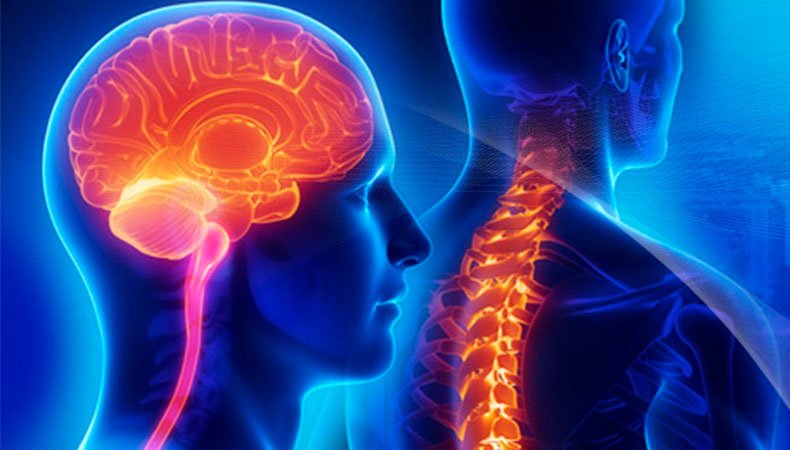
Brain and Spine tumours
| Dr Vishakha
Tumours are abnormal tissue growths caused by cells growing out of control. They can start in the brain or spine, called primary tumours, or spread through the bloodstream, called metastatic tumours. These rare tumours affect about 25,000 people, with only 1% of the population having a brain tumour in their lifetime. The most common types of brain tumours are metastatic and require expert treatment. Common cancers that spread to the brain include lung, breast, melanoma, and colon cancer. Spine tumours compress the spinal cord and nerves, making treatment complex and requiring evaluation by experienced neurosurgeons and oncologists.

Brain tumours:
Gliomas:
- These include glioblastoma multiforme (GBM), astrocytoma, and oligodendrogliomas.
- Meningiomas:
- Arise from the meninges, the layers covering the brain.
- Pituitary tumours:
- Develop in the pituitary gland at the base of the brain, affecting hormone production.
- Medulloblastoma:
- Children’s cerebellum is standard.
- Schwannomas:
- They are originating from Schwann cells, often affecting vestibular and auditory nerves.
- Craniopharyngioma:
- Due to its location near the pituitary gland and hypothalamus, it is often diagnosed in children.
- Pineal region tumours:
- Examples include pineocytomas and pineoblastomas.
- Chordomas:
- Arise from notochord remnants, commonly found in the base of the skull or along the spine.
Spine tumours:
- Ependymomas:
- Originate from ependymal cells that line the spinal cord’s central canal.
- Astrocytomas:
- Similar to their brain counterparts, astrocytomas in the spinal cord are rare but can occur.
- Schwannomas:
- Also known as neurilemmomas, these tumours arise from Schwann cells in peripheral nerves of the spinal cord.
- Meningiomas:
- Meningiomas can also occur in the spinal cord, typically in the thoracic region.
- Hemangioblastomas:
- Found in the cervical and thoracic spinal cord, associated with von Hippel-Lindau (VHL) disease.
- Lipomas:
- The spinal cord or filum terminale may develop benign fatty tumours.
- Germ cell tumour:
- Rare tumours occur in the pineal or suprasellar regions, indirectly affecting the spinal cord.
- Intradural extramedullary tumours:
- Develop within the protective covering of the spinal cord but outside the spinal cord itself.
Causes and symptoms of brain and spine tumours:
Causes of brain tumour:
- Age:
- Brain tumours can occur at any age, but certain types are more common in specific age groups.
- For instance, medulloblastomas are more common in youngsters, whereas gliomas are more common in adults.
- Genetics:
- Some individuals may have a genetic predisposition to developing certain types of brain tumours.
- Genetic syndromes, such as neurofibromatosis, von Hippel-Lindau (VHL) syndrome, and Li-Fraumeni syndrome, increase the risk.
- Radiation exposure:
- Exposure to ionizing radiation, especially at a young age, is a known risk factor for certain brain tumours.
- It includes radiation therapy for the treatment of other cancers.
- Family history:
- A family history of brain tumours may increase an individual’s risk, suggesting a possible genetic component.
- Immune system conditions:
- Some conditions that affect the immune system, such as HIV/AIDS, may be associated with an increased risk of certain brain tumours.
- Environmental factors:
- Exposure to certain environmental toxins or chemicals has been studied as a potential risk factor, but the evidence often needs to be more conclusive.
- Previous:
- Individuals who have undergone radiation therapy for previous cancers, especially in the head and neck area, may have an increased risk.
Symptoms of brain tumour:
- Headache:
- Persistent or severe headaches, often worse in the morning or with changes in position
- Seizures:
- Epilepsy patients with new seizures or altered seizure patterns.
- Neurological symptoms:
- Limb weakness and numbness are usually confined to one side of the body.
- Changes in coordination or balance.
- Difficulty walking or unexplained falls.
- Vision changes:
- Blurred or double vision.
- Loss of peripheral vision impairments.
- Speech and language difficulties:
- Changes in speech, including slurred words
- Needs help in pronouncing words correctly.
- Difficulty in walking:
- Problems with balance and walking can be related to the tumour’s location.
- Changes in sensation:
- Changes in sensation, such as tingling or numbness, may occur in the extremities.
- Behavioural changes:
- A change of personality, a mood swing, or a change of behaviour.
- Hormonal changes:
- Pituitary tumours can cause hormonal imbalances, leading to symptoms such as irregular menstruation, growth abnormalities, or changes in sexual function.
- Cognitive changes:
- Memory loss with difficulty concentration.
- Personality changes or alterations in mood.
- Nausea and vomiting:
- Unexplained nausea and vomiting, often accompanied by headaches.
Various conditions can cause symptoms of neurological issues and do not necessarily indicate a brain tumour. However, if an individual experiences persistent or worsening neurological symptoms, they should seek medical attention for a thorough evaluation and diagnosis. Early detection and intervention are crucial for better outcomes in the treatment of brain tumours. It is important to note that various conditions can cause these symptoms and do not necessarily indicate a brain tumour.
Causes of spine tumour:
- Primary tumours:
- Intramedullary tumours: The term “arise” refers to the process of forming within the spinal cord’s cells.
- Extramedullary tumours: The development of the spinal cord occurs in the supporting structures around it, including the meninges, nerve roots, and vertebrae.
- Metastatic tumours:
- Cancer cells from other parts of the body can metastaticize and spread to the spine.
- Genetic factors:
- Neurofibromatosis is a genetic condition that can heighten the likelihood of developing spinal tumours.
- Age:
- Certain age groups are more likely to develop certain types of spinal tumours.
- Radiation exposure:
- Previous radiation exposure, either for medical treatment or other reasons, may increase the risk of developing spinal tumours.
- Immunosuppression:
- Conditions that weaken the immune system, such as HIV/AIDS or organ transplantation, may be associated with an increased risk of certain tumours.
Symptoms of spine tumours:
- Back pain:
- Persistent and often severe back pain that may worsen at night or with movement.
- Radicular pain:
- Pain radiates along the nerves, which may extend into the arms or legs.
- Weakness and numbness:
- Weakness or numbness in the limbs, potentially leading to difficulty walking or performing delicate motor tasks.
- Changes in bowels or bladder dysfunction:
- Incontinence or trouble managing bladder or bowel movements.
- Sensory changes:
- Changes in sensation, such as tingling or loss of sensation in specific areas of the body.
- Difficulty in walking:
- Problems with balance and coordination.
- Spinal deformity:
- In some cases, the tumour may cause changes in the alignment of the spine.
- Muscle spasms:
- Spasms or cramping in the muscles of the back or neck.
- Difficulty with coordination:
- Lack of coordination and difficulty with fine motor skills.
- Spinal cord compression:
- Compression of the spinal cord can lead to a range of symptoms, including pain, weakness, and changes in sensation.
Various conditions can cause symptoms of spinal tumours and do not necessarily indicate the presence of a tumour. Prompt medical evaluation is crucial for a thorough diagnosis, and diagnostic tests like MRI and CT scans are used to confirm the presence and type of the tumour. Early detection and appropriate intervention are crucial to managing spinal tumours and improving outcomes.
Test recommendations for brain tumour diagnosis:
Brain tumour diagnosis involves tests to assess tumour location, size, type, and characteristics, with specific tests based on patient symptoms, medical history, and suspected tumour type, requiring careful examination.
- Neurological examination:
- A thorough assessment of neurological function, including reflexes, strength, coordination, and sensory perception, to identify any abnormalities.
- Imaging studies:
- MRI(Magnetic resonance imaging):
- Produces detailed images of the brain, allowing visualization of the tumour’s location, size, and characteristics.
- CT(Computed Tomography)scan:
- It provides cross-sectional images and helps detect abnormalities, including tumours.
- It may be used in conjunction with MRI.
- Biopsy:
- Stereotactic biopsy:
- In cases where the tumour is deep or complex to access, a stereotactic biopsy may be performed using specialized imaging guidance to obtain a small tissue sample for analysis.
- Open biopsy:
- In some cases, particularly during surgical procedures, a larger tissue sample may be obtained for examination.
- Cerebrospinal fluid(CSF)examination:
- A lumbar puncture (spinal tap) may be performed to analyze the cerebrospinal fluid for the presence of cancer cells, blood, or other abnormalities.
- Angiography:
- A study of the blood vessels in and around the brain using contrast material.
- It helps assess blood flow and detect abnormalities related to certain types of brain tumours.
- Positron emission tomography(PET) scan:
- Measures metabolic activity in tissues and can help differentiate between benign and malignant tumours.
- PET scans are often used in conjunction with other imaging studies.
- Electroencephalogram(EEG):
- Records the electrical activity of the brain, helping to identify abnormal patterns associated with seizures or certain types of brain tumours.
- Functional MRI(fMRI):
- It maps brain activity and may be used to identify critical areas involved in functions such as movement, language, and sensory perception.
- It assists in surgical planning to avoid damage to vital brain regions.
- Genetic testing:
- In some cases, genetic testing may be recommended, especially if there is a family history of certain genetic conditions associated with an increased risk of brain tumours.
- Visual field testing:
- Assesses changes in peripheral vision, which can be affected by certain tumours, particularly those involving the optic nerve or optic chiasm.
The combination of tests for brain tumour diagnosis depends on the patient’s circumstances and suspected tumours. Collaboration among neurologists, neurosurgeons, oncologists, and radiologists is crucial for accurate diagnosis and treatment planning, ensuring timely and effective interventions for brain tumours.
Test recommendations for spine tumour diagnosis:
Spinal tumour diagnosis involves clinical evaluations, imaging studies, and biopsy procedures, with specific tests based on patient symptoms, medical history, and suspected tumour type and location.
- Neurological examination:
- A thorough assessment of neurological function, including reflexes, strength, coordination, and sensory perception, to identify any abnormalities related to the spinal cord and nerve roots.
- Clinical history and physical examination:
- A complete evaluation of the patient’s medical history, encompassing the genesis and advancement of symptoms, succeeded by a physical assessment aimed at assessing neurological function, reflexes, and indications of spinal cord compression.
- Imaging studies:
- MRI(Magnetic resonance imaging):
- Provides detailed images of the spinal cord and surrounding structures, helping to visualize the location, size, and characteristics of the tumour.
- CT(computed tomography)scan:
- It offers cross-sectional images helps detectprovideormalities and provides additional information about the tumour.
- Biopsy:
- Needle biopsy:
- In cases where the tumour is accessible, a needle biopsy may be performed under imaging guidance to obtain a small tissue sample for analysis.
- Open biopsy:
- In some cases, particularly during surgical procedures, a larger tissue sample may be obtained for examination.
- Cerebrospinal fluid(CSF)examination:
- A lumbar puncture (spinal tap) may be performed to analyze the cerebrospinal fluid for the presence of cancer cells or other abnormalities, particularly when the tumour is affecting the spinal cord.
- Myelogram:
- It involves injecting contrast dye into the spinal canal and performing a CT scan.
- It helps visualize the spinal cord and nerve roots, mainly when other imaging studies may not provide sufficient information.
- Electromyography(EMG) and nerve conduction studies:
- Measures electrical activity in muscles and nerves, helping to assess nerve function and detect abnormalities related to spinal cord compression.
- Angiography:
- An imaging technique that uses contrast material to visualize blood vessels in and around the spine.
- It may be employed to assess blood flow and detect abnormalities related to certain types of spinal tumours.
- Positron emission tomography(PET) scan:
- Determines the metabolic activity of tissues, offering more details about a tumour’s properties and possible routes of dissemination.
- Genetic testing:
- In some instances, genetic testing may be recommended, especially if there is a family history of genetic conditions associated with an increased risk of spinal tumours.
- Bone scan:
- A nuclear medicine test that helps identify abnormalities in the bones helps detect metastatic tumours in the spine.
The combination of tests for spinal tumour diagnosis depends on the patient’s circumstances and the nature of the suspected tumour. A collaborative approach involving neurologists, surgeons, oncologists, and radiologists is crucial for accurate diagnosis and effective treatment, ensuring timely and precise interventions.
The primary treatment modalities for brain and spine tumours include:
The treatment of brain and spine tumours is determined by factors like tumour type, location, size, grade, patient’s health, and spread. A multidisciplinary team, including neurologists, neurosurgeons, oncologists, and radiation oncologists, collaborates to develop an individualized treatment plan.
- Surgery:
The goal is to remove the tumour if it is possible.
- Radiation therapy:
- The use of targeted radiation is used to either eliminate or control tumour cells.
- Chemotherapy:
- Systemic drugs are used to eradicate cancer cells.
- Targeted therapies:
- Drugs that target specific molecular pathways in cancer cells.
- Stem cell transplant:
- This term is utilized in certain instances, especially for aggressive tumours.
Prognosis:
- Varies:
- The outcome of a tumour depends on its type, location, and grade, as well as the patient’s overall health.
- Early detection:
- Increases the chance that the treatment will be successful.
- Multidisciplinary approach:
- The process involves involving specialists for comprehensive care.
Supportive care:
- Rehabilitation:
- Physical therapy is being utilized to restore functional abilities.
- Pain management:
- The focus is on managing the pain linked to the tumour.
- Psychological support:
- We are managing the emotional and psychological aspects of the diagnosis.
Supportive care is crucial for children with brain and spine tumours, involving a multidisciplinary approach involving pediatric oncologists, neurosurgeons, and radiation oncologists. It manages treatment side effects, provides psychological support, and addresses the long-term effects of cancer. Early diagnosis and treatment advancements have improved the outlook for many children.
Proficiency of Dr Vishaka:
Hydrocephalus (increased fluid in the brain): The procedure involves an endoscopic third ventriculostomy and CSF diversion (VP shunt) to treat complex hydrocephalus.
- Craniosynostosis (abnormal head shape due to untimely cranial sutures fusion) surgeries: Helmet therapy is a technique that is used in both endoscopic and open surgery.
- Spinal dysraphisms(Spina Bifida)- (spinal abnormalities present by birth) – surgical repair
- Encepahaocles repair surgery.
- Vascular conditions and stroke surgeries: revascularization surgeries for moya moya disease.
- Pediatric brain and spine tumour surgeries.
- Pediatric brain and spine infection surgeries: Endoscopic and open surgeries for brain and spine infections.
- Pediatric traumatic brain and spinal injury.
- Antenatal counselling for congenital fatal neurosurgical conditions.
Dr Vishaka specializes in craniosynostosis surgery, which is only done in a few centres in India. Dr Vishaka Patil, M.B.B.S, DNB (AIIMS) New Delhi, M.Ch (IPGMER SSKM) became a Member of “The Royal College of Surgeons, Edinburgh” (U.K.) a highly successful and best pediatric neurosurgeon in Hyderabad, Telangana with 13 years of experience, is among the topmost pediatric neurosurgeons in the Rainbow group of hospitals at Hyder Nagar and Banjara Hills.

