Encephalocele Repair

An overview of Encephalocele repair:
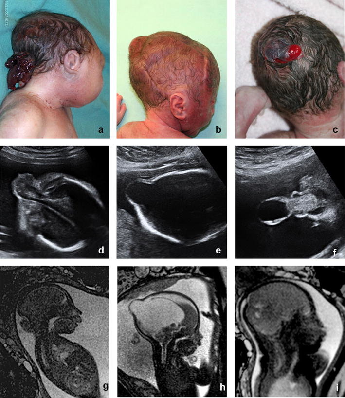
Encephalocele is a neural tube abnormality that results in a sac-like protrusion of the brain and skull membranes owing to inadequate neural tube closure during foetal development.
Types of Encephalocele repair:
- Frontal encephalocele: Frontal encephalocele is a rare neural tube defect involving a sac-like protrusion containing brain tissue and membranes, extending through an opening in the frontal part of the skull.
- Occipital encephalocele: This is a common neural tube defect characterised by a sac-like protrusion of brain tissue and meninges that extends through an incision in the occipital area of the skull.
- Perinatal encephalocele: Parietal encephalocele is a rare neural tube defect involving a sac-like protrusion containing brain tissue and meninges that extends through an opening in the upper middle part of the skull.
- Basal encephalocele: Parietal encephalocele is a rare neural tube defect involving a sac-like protrusion containing brain tissue and meninges that extends through an opening in the upper middle part of the skull.
Purpose of encephalocele repair:
The treatment involves sealing the skull defect, removing non-functional brain tissue, covering the brain with healthy tissues or synthetic materials, and improving neurological function by addressing and alleviating symptoms associated with the encephalocele.
Treatment for encephalocele:
The process involves repairing the skull defect, covering it with normal tissues, removing herniated brain tissue, and preventing complications like infections and hydrocephalus.
Surgical procedure of encephalocele repair:
The patient undergoes general anaesthesia, followed by an incision to expose the encephalocele. The herniated brain tissue is carefully repositioned or excised, and the skull defect is repaired using bone grafts, synthetic materials, or a combination of both. The incision is closed in layers to promote healing while minimising infection and cerebrospinal fluid leakage.
Postoperative care for encephalocele repair:
Intensive monitoring involves close monitoring in a care setting for complications like infection, increased pressure, and cerebrospinal fluid leaks. Medications include antibiotics for infection prevention and pain management. Rehabilitation includes physical, occupational, and developmental support for recovery.
Recovery time for encephalocele repair:
An encephalocele’s healing duration is impacted by its size and location, the existence of hydrocephalus, and other congenital defects or genetic diseases. Early diagnosis and timely surgical intervention improve recovery outcomes. Physical therapy, occupational therapy, and developmental support are initiated to address neurological or developmental delays. Gradual return to school and normal activities depend on the child’s health and recovery needs. Regular follow-up visits are conducted to monitor neurological function, development, and surgical repair integrity, as well as to address any complications.
Prognosis:
The encephalocele’s size and location, the presence of Hydrocephalus, and associated anomalies can impact its prognosis. Early diagnosis and prompt surgical intervention can improve outcomes, while other congenital anomalies or genetic syndromes can also influence the outcome.
Encephalocele repair is a complex surgical procedure requiring early detection, appropriate intervention, and comprehensive postoperative care. Successful management requires multidisciplinary care involving neurosurgeons, paediatricians, and rehabilitation specialists, aiming for improved quality of life for affected individuals.
About Dr Vishakha
Dr. Vishakha Karpe, a highly skilled Senior Paediatric Neurosurgeon at Rainbow Children’s Hospital, Banjara Hills, and Hyder Nagar in Hyderabad, is one of India’s leading paediatric neurosurgeons with extensive experience in paediatric neurosurgery. With over nine years of dedicated practice, she is among the few in India working extensively in this field.
With extensive experience in paediatric neurosurgical conditions, she focuses on comprehensive care, including precise surgery and educating parents about the complete case management protocol. She is an efficient and passionate medical professional, pursuing ethical practice and ensuring patient care after surgery.
Proficiency of Dr Vishaka:
- Hydrocephalus (increased fluid in the brain) Treatment:
Endoscopic third ventriculostomy and CSF diversion (VP shunt) , neuroendoscopic
procedures for complex hydrocephalus. - Craniosynostosis (abnormal head shape due to untimely cranial sutures fusion) :
- Treatment –
Helmet therapy , endoscopic and open surgery. - Spinal dysraphisms(Spina Bifida)-
(spinal abnormalities present since birth) – surgical repair - Encephalocele repair surgery.
- Vascular and stroke surgeries:
revascularization surgeries for moya moya disease, emergency surgery for stroke - Pediatric brain and spine tumour surgeries.
- Pediatric brain and spine infection surgeries:
Endoscopic and open surgeries for brain and spine infections. - Pediatric traumatic brain and spinal injury.
- Antenatal counselling for congenital fatal neurosurgical conditions.
Dr Vishakha Karpe M.B.B.S, DNB (AIIMS) New Delhi, M.Ch (IPGMER SSKM) Member of “The Royal College of Surgeons, Edinburgh” (U.K.) is a highly competent and one of the best pediatric neurosurgeons in Hyderabad, Telangana with 13 years of experience, is among the topmost pediatric neurosurgeons in the Rainbow group of hospitals at Banjara Hills and Hyder Nagar.

