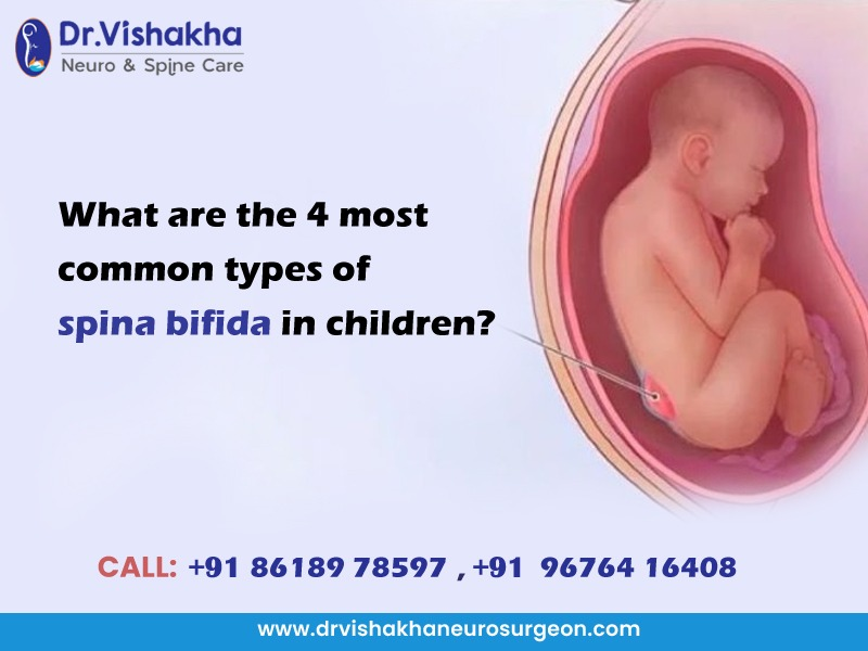
Spina bifida is a congenital condition caused by incomplete neural tube closure during early fetal development. It affects the spine and spinal cord, causing mild to severe physical and neurological impairments. This essay explores the types, causes, diagnosis, treatment, and prevention of spina bifida, emphasising its impact on affected individuals and the importance of early intervention.
Classifications of Spina Bifida
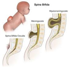
- Spine Bifida Occulta
Spina bifida occulta is the mildest form of spina bifida, a neural tube defect during early fetal development. It is characterised by a small vertebral defect where bones fail to close around the spinal cord. Unlike more severe forms, the spinal cord and surrounding nerves remain intact.
Causes and symptoms
Spina bifida occulta is a condition resulting from incomplete neural tube closure during embryonic development, usually between the third and fourth weeks of pregnancy. Contributing factors include genetic predisposition, environmental factors like folate deficiency, maternal diabetes or obesity, and exposure to certain medications or toxins.
Spina bifida occulta is a condition where the spinal cord is tethered, causing a condition where the spinal cord is tethered to the body. Symptoms can include a small dimple, birthmark, hair tuft, or slight skin indentation. In rare cases, it may cause back pain, leg weakness, bladder or bowel dysfunction, or worsening neurological symptoms with growth.
Complication and prevention
Spina bifida occulta can cause tethered spinal cord syndrome. In this condition, the spinal cord becomes abnormally attached to surrounding tissues, resulting in neurological deficits, syringomyelia, a cyst within the spinal cord, and orthopaedic issues like scoliosis or foot deformities.
Spina bifida occulta can be reduced during pregnancy by consuming 400-800 micrograms of folic acid daily from leafy greens, fortified cereals, beans, and citrus fruits. Prenatal care includes regular medical check-ups and ultrasounds, as well as avoiding harmful substances like alcohol, tobacco, and certain medications.
- Meningocele
Meningocele is a type of spina bifida, a neural tube defect where the spinal column fails to close completely during fetal development. It is a moderate form of spina bifida involving the protrusion of the meninges through an opening in the spine. This condition reduces the likelihood of severe neurological deficits by leaving the spinal cord intact. Meningocele is characterized by a sac-like protrusion on the back, containing cerebrospinal fluid and meninges but no spinal cord tissue.
Causes and symptoms
Meningocele is caused by a combination of genetic and environmental factors, including folate deficiency during pregnancy, genetic predisposition with neural tube defects, maternal health conditions like diabetes or obesity, and exposure to certain medications or harmful substances during pregnancy.
Meningocele symptoms vary based on the defect’s location and size. Typically, a fluid-filled sac on the back, usually in the mid to lower spine, is visible. Neurological function is usually preserved, but mild motor or sensory impairments may occur if there are tethering or other abnormalities. Other potential issues include the risk of infection if the sac ruptures and potential bladder or bowel dysfunction, depending on the defect’s location.
Complications
Meningocele can cause infections, neurological deficits, bladder or bowel dysfunction, and hydrocephalus if untreated or improperly managed. The exposed sac is vulnerable to infections like meningitis, and other spinal anomalies may affect related nerve roots. Disruption of cerebrospinal fluid circulation can also lead to hydrocephalus.
Prevention
Prenatal health and nutrition are prioritized, with women consuming 400-800 micrograms of folic acid daily from fortified cereals, leafy greens, beans, and citrus fruits. Regular medical check-ups and ultrasounds are also recommended, along with avoiding harmful substances like alcohol and tobacco. Pre-existing conditions like diabetes and obesity can be managed.
- Myelomeningocele
Myelomeningocele is a severe and complex form of spina bifida, a neural tube defect caused by improper formation of the spine and spinal cord during early fetal development. It involves the protrusion of the spinal cord, meninges, and cerebrospinal fluid through an open vertebrae defect, forming a visible sac on the back. This condition often results in significant neurological and physical impairments, typically occurring in the lumbar or sacral regions, but can affect any part of the spine.
Causes and symptoms
Myelomeningocele is a genetic condition linked to insufficient maternal folate intake during pregnancy, a family history of neural tube defects, and environmental factors such as maternal obesity, diabetes, exposure to certain medications, and exposure to high temperatures during early pregnancy. The exact cause is not fully understood.
The severity of symptoms depends on the defect’s location and size. Common symptoms include a sac-like protrusion on the back containing spinal cord tissue, meninges, and cerebrospinal fluid. Neurological impairments include weakness or paralysis, loss of sensation in the lower body, bladder and bowel dysfunction, and tethered cord syndrome. Associated conditions include hydrocephalus, Chiari Malformation Type II, and orthopaedic problems like scoliosis, clubfoot, hip dislocation, or joint contractures. Treatment with a shunt is common in 70-90% of cases.
Complications
Myelomeningocele can cause various complications, including infections, neurological damage, hydrocephalus, bladder and bowel problems, learning disabilities, and mobility challenges. Open defects increase the risk of meningitis, while hydrocephalus increases intracranial pressure, requiring shunting. Bladder and bowel problems can lead to lifelong incontinence or retention issues.
Preventions
To prevent myelomeningocele, women should supplement with folic acid and include fortified foods in their diet. Prenatal care should include regular check-ups, ultrasounds, and early screening for neural tube defects. Teratogens should be avoided, and maternal conditions should be managed, including controlling diabetes and obesity before and during pregnancy.
- Closed Neural Tube Defects
Closed neural tube defects (CNTDs) are a type of spina bifida characterized by malformations in the spinal cord, vertebrae, or surrounding tissues covered by skin. These defects are not exposed to the body, reducing the risk of infection and severe neurological damage. However, they can still cause varying degrees of neurological and physical symptoms depending on the severity and location of the defect. CNTDs occur during early embryonic development.
Types of closed neural tube defects
- Lipomyelomeningocele is a fatty mass that can bind to the spinal cord, potentially causing neurological issues.
- Lipomeningocele is a condition where a fatty mass protrudes through a vertebral opening while the spinal cord remains in its normal position.
- The tethered spinal cord, an abnormally attached part of the spinal cord, can restrict movement and cause stretching during child growth.
- Diastematomyelia is a condition where the spinal cord is divided into two parts, often separated by a bony or fibrous band.
- The Dermal Sinus Tract is a narrow channel from the skin to the spinal cord or its coverings, increasing the risk of infection.
- Sacral agenesis refers to the partial or complete absence of the sacrum, the lower part of the spine, often linked to other malformations.
Causes and symptoms
The cause of CNTDs is not fully understood but is influenced by genetic and environmental factors. Genetic predisposition includes a family history of neural tube defects, folic acid deficiency during pregnancy, maternal diabetes, obesity, exposure to teratogens, and environmental influences like high temperatures during early pregnancy.
CNTDs can present with various symptoms depending on the type and severity of the defect. Visible signs include a dimple, birthmark, fatty mass, or small opening. Neurological symptoms include leg weakness, loss of sensation, bladder or bowel dysfunction, and orthopaedic problems like scoliosis, clubfoot, joint contractures, or hip dislocation. Other symptoms may include asymptomatic cases or more severe conditions.
Complications
Closed neural tube defects can cause various complications, such as tethered spinal cord syndrome, neurological damage, bladder and bowel dysfunction, infections, and orthopaedic issues, such as scoliosis, foot deformities, and gait abnormalities. These complications can result in progressive neurological deficits, weakness, numbness, and loss of motor function.
Prevention
To reduce neural tube defects during pregnancy, women should consume 400-800 micrograms of folic acid daily, starting one month before conception and continuing through the first trimester. Healthy pregnancy practices include regular prenatal check-ups, avoiding harmful substances, managing chronic conditions like diabetes and obesity, and promoting awareness about the importance of folic acid and prenatal care.
Diagnosis
Spina bifida can be diagnosed prenatally or after birth through screening tests like maternal serum alpha-fetoprotein (AFP) testing and ultrasound imaging. Amniocentesis confirms the diagnosis by analyzing AFP and acetylcholinesterase levels in amniotic fluid. Postnatal diagnosis involves physical examination and imaging studies like MRI or CT scans to evaluate spinal and neurological involvement.
Treatment
Spina bifida is a complex condition that requires multidisciplinary treatment. Postnatal surgery is typically performed within 48 hours of birth to prevent infections and treat hydrocephalus. Complications include bladder and bowel dysfunction through clean intermittent catheterization, medications, or reconstructive surgeries. Orthopaedic issues are addressed through physical therapy, bracing, or corrective surgeries. Rehabilitation is crucial for improving mobility, strength, and independence. In cases of myelomeningocele, fetal surgery may be performed to reduce neurological complications.
Prognosis
Spina bifida’s prognosis depends on the defect’s type, severity, and complications. Mild forms can live without symptoms, while severe forms like myelomeningocele can lead to lifelong challenges like mobility limitations, bladder and bowel dysfunction, and infection risk. Advances in surgical and supportive care have improved survival rates and quality of life, allowing many to lead fulfilling lives.
Spina bifida is a lifelong condition with significant medical, physical, and social implications. Early diagnosis, advanced surgical techniques, and multidisciplinary care have improved outcomes. Public health measures, particularly folic acid supplementation, are crucial in reducing the incidence. Research and advocacy are needed for better treatments, support, and opportunities for individuals with spina bifida.
About Dr Vishakha
Dr. Vishakha Karpe, a highly skilled Senior Paediatric Neurosurgeon at Rainbow Children’s Hospital, Banjara Hills, and Hyder Nagar in Hyderabad, is one of India’s leading paediatric neurosurgeons with extensive experience in paediatric neurosurgery. With over nine years of dedicated practice, she is among the few in India working extensively in this field.
With extensive experience in paediatric neurosurgical conditions, she focuses on comprehensive care, including precise surgery and educating parents about the complete case management protocol. She is an efficient and passionate medical professional, pursuing ethical practice and ensuring patient care after surgery.
Proficiency of Dr Vishaka:
Hydrocephalus (increased fluid in the brain): The procedure involves an endoscopic third ventriculostomy and CSF diversion (VP shunt) to treat complex hydrocephalus.
- Craniosynostosis (abnormal head shape due to untimely cranial sutures fusion) surgeries: Helmet therapy is a technique that is used in both endoscopic and open surgery.
- Spinal dysraphisms(Spina Bifida)- (spinal abnormalities present by birth) – surgical repair
- Encepahaocles repair surgery.
- Vascular conditions and stroke surgeries: revascularization surgeries for moya moya disease.
- Pediatric brain and spine tumour surgeries.
- Pediatric brain and spine infection surgeries: Endoscopic and open surgeries for brain and spine infections.
- Pediatric traumatic brain and spinal injury.
- Antenatal counselling for congenital fatal neurosurgical conditions.
Dr Vishaka specializes in craniosynostosis surgery, which is only done in a few centres in India. Dr Vishaka Patil, M.B.B.S, DNB (AIIMS) New Delhi, M.Ch (IPGMER SSKM) became a Member of “The Royal College of Surgeons, Edinburgh” (U.K.) a highly successful and best pediatric neurosurgeon in Hyderabad, Telangana with 13 years of experience, is among the topmost pediatric neurosurgeons in the Rainbow group of hospitals at Hyder Nagar and Banjara Hills.