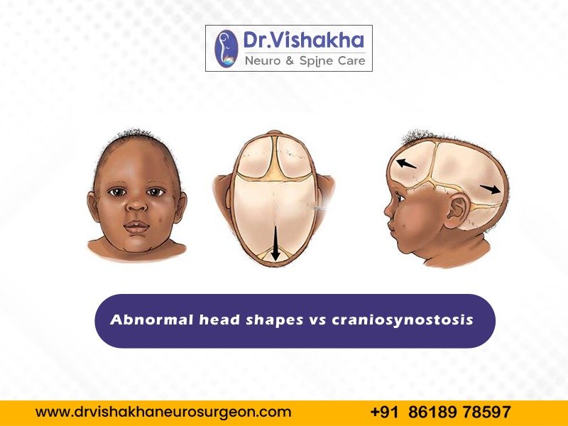
Abnormal head shapes in infants and children can be caused by various factors, including positional issues and developmental issues, and understanding these conditions is crucial for proper diagnosis and treatment.
The primary causes and reasons for unusual head shapes include:
- Positional factors:
- Positional Plagiocephaly: Long periods of sleeping in one position are a common cause of back pain in infants, particularly during the first few months. Prevention involves frequent position changes, supervised tummy time, and helmet use if necessary.
- Positional Brachycephaly: Prolonged back laying flattens the head due to occiput pressure. Prevention/treatment involves alternating positions and supervised tummy time, similar to positional plagiocephaly.
- Birth trauma: The cause of this condition is the pressure during delivery, particularly prolonged labour or instrumental deliveries, which can temporarily mould or despise the soft skull bones. Treatment typically self-corrects over time, but monitoring and physical therapy may be necessary.
- Torticollis: Tight neck muscles cause a baby’s head to tilt, leading to uneven pressure on the skull. Physical therapy to strengthen and stretch the neck muscles is a preventative measure.
- Premature birth: Prematurity causes softer skulls in premature infants, leading to prolonged NICU stays. Prevention involves careful repositioning and supportive devices to minimise pressure on the skull.
- Congenital conditions: Premature fusion of cranial sutures due to genetic factors or mutations restricts skull growth in certain areas. Treatment requires surgical intervention to correct skull shape and allow average brain growth.
- External pressures: Premature fusion of cranial sutures due to genetic factors or mutations restricts skull growth. Preventative treatment requires surgical intervention to correct skull shape and average brain growth. Genetic syndromes like Apert and Crouzon syndrome often involve multiple anomalies, affecting skull and facial development. Multidisciplinary medical management, including surgery and genetic counselling, is recommended for prevention and treatment.
- Environmental and cultural practices: Swaddling or cradling practices can cause prolonged pressure on specific head parts, and prevention involves educating caregivers about culturally sensitive practices and varying baby head positions.
Most common abnormal head shapes:
- Plagiocephaly: Plagiocephaly is a skull disorder characterized by asymmetrical distortion of one side. Positional Plagiocephaly occurs when a baby lies in one position for extended periods and is usually corrected with positioning changes and physical therapy. Craniosynostosis causes congenital plagiocephaly, which is uncommon.
- Brachycephaly: This flattened head appearance, often with asymmetry of ears, forehead, and face, is caused by positional factors, womb positioning, or external pressures. Treatment options include repositioning techniques, physical therapy, and helmet therapy.
- Scaphocephaly (Dolichocephaly): The head is long and narrow, caused by premature sagittal suture fusion or positional issues, particularly in premature infants in the NICU, and can be treated with surgery or repositioning.
- Trigonocephaly: Trigonocephaly is a type of craniosynostosis with a triangular-shaped forehead due to premature fusion of the metopic suture. This closure restricts frontal bone growth and affects the forehead’s shape and brain space. The primary cause is an early closure of the metopic suture that connects the crown of the head to the nose.
What is Craniosynostosis?
Craniosynostosis is a congenital disability where the fibrous joints between a baby’s skull close prematurely, preventing average skull growth and potentially causing a misshapen head and developmental issues.
Types of Craniosynostosis:
- Sagittal synostosis (Scaphocephaly): Scaphocephaly is the most common type of craniosynostosis, defined as a long and narrow head shape caused by premature fusion of the sagittal suture.
- Coronal synostosis: Premature fusion of coronal sutures results in an asymmetrical head shape (anterior plagiocephaly) or broad, short head (brachycephaly) due to Unicoronal synostosis, running from ear to ear over the head.
- Metopic synostosis: Premature metopic suture fusion, running from head top to nose, with triangular forehead, ridge, and close-set eyes.
- Lambdoid synostosis: An untimely lambdoid suture combination on the head back straightens the influenced side’s back, coming about in back plagiocephaly.
- Multiple suture synostosis: Premature fusion of numerous sutures produces a complex and variable head shape that is frequently associated with syndromic forms of craniosynostosis.
Causes and symptoms of Craniosynostosis
Genetic mutations in genes like FGFR1, FGFR2, FGFR3, and TWIST, syndromic conditions like Apert syndrome, Crouzon syndrome, Pfeiffer syndrome, and Muenke syndrome, and environmental factors like maternal smoking can cause craniosynostosis.
Abnormal head shape, ridge along the fused suture, cognitive and developmental delays due to increased intracranial pressure, facial asymmetry, and vision problems are common side effects of certain sutures, particularly coronal or metopic sutures, and can be influenced by abnormal skull growth.
Abnormal head shape v/s Craniosynostosis:
| Abnormal head shape | Craniosynostosis |
| Abnormal head shape, external pressures and positioning, and premature suture fusion. | External pressures, positioning, and premature suture fusion are all common causes of craniosynostosis. |
| Abnormal head shapes can be corrected with repositioning and physical therapy. | Craniosynostosis requires surgical intervention to correct skull shape and promote average brain growth. |
| Craniosynostosis is a condition in which cranial sutures close prematurely. | Skull growth is restricted in one area while increasing in others, resulting in an abnormal head shape. |
Positional head shape abnormalities in children typically respond well to non-surgical treatments without long-term effects. For craniosynostosis, early surgical intervention leads to good outcomes and ongoing monitoring for normal development. Craniosynostosis is treatable and generally favourable when diagnosed and managed early. Early surgical intervention prevents complications and ensures normal brain and skull development. If suspected, prompt evaluation by a specialist is essential for the best outcomes. Understanding the differences between positional factors and craniosynostosis is crucial for proper diagnosis and management. Addressing abnormal head shapes early is necessary for optimal outcomes for affected children.
About Dr Vishakha
Dr. Vishakha Karpe, a highly skilled Senior Paediatric Neurosurgeon at Rainbow Children’s Hospital, Banjara Hills, and Hyder Nagar in Hyderabad, is one of India’s leading paediatric neurosurgeons with extensive experience in paediatric neurosurgery. With over nine years of dedicated practice, she is among the few in India working extensively in this field.
With extensive experience in paediatric neurosurgical conditions, she focuses on comprehensive care, including precise surgery and educating parents about the complete case management protocol. She is an efficient and passionate medical professional, pursuing ethical practice and ensuring patient care after surgery.
Proficiency of Dr Vishaka:
Hydrocephalus (increased fluid in the brain): The procedure involves an endoscopic third ventriculostomy and CSF diversion (VP shunt) to treat complex hydrocephalus.
- Craniosynostosis (abnormal head shape due to untimely cranial sutures fusion) surgeries: Helmet therapy is a technique that is used in both endoscopic and open surgery.
- Spinal dysraphisms(Spina Bifida)- (spinal abnormalities present by birth) – surgical repair
- Encepahaocles repair surgery.
- Vascular conditions and stroke surgeries: revascularization surgeries for moya moya disease.
- Pediatric brain and spine tumour surgeries.
- Pediatric brain and spine infection surgeries: Endoscopic and open surgeries for brain and spine infections.
- Pediatric traumatic brain and spinal injury.
- Antenatal counselling for congenital fatal neurosurgical conditions.
Dr Vishaka specializes in craniosynostosis surgery, which is only done in a few centres in India. Dr Vishaka Patil, M.B.B.S, DNB (AIIMS) New Delhi, M.Ch (IPGMER SSKM) became a Member of “The Royal College of Surgeons, Edinburgh” (U.K.) a highly successful and best pediatric neurosurgeon in Hyderabad, Telangana with 13 years of experience, is among the topmost pediatric neurosurgeons in the Rainbow group of hospitals at Hyder Nagar and Banjara Hills.


