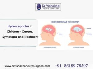
- Understanding Infant Skull Development
- The newborn skull consists of multiple bones (frontal, parietal, occipital, temporal) joined by cranial sutures and fontanelles.
- These flexible joints allow brain growth and birth passage.
- Head shape variations are common in the first few months due to moulding during delivery and sleeping posture.
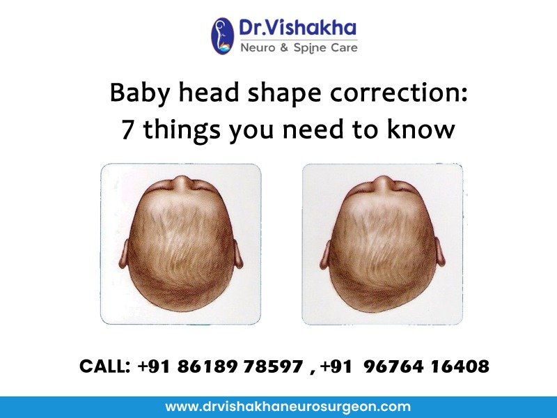
- What is Positional Plagiocephaly?
- The most frequent cause of an abnormal head shape is.
- Characterised by flattening on one side of the occiput, often due to consistent supine sleeping position.
- Difference from craniosynostosis (a pathological condition where sutures close prematurely).
- Identifying When Head Shape Needs Medical Attention
Red flags:
Asymmetry that worsens despite repositioning.
Abnormal ridges over sutures.
Delayed or abnormal head growth on percentile charts.
A paediatrician or neurosurgeon may recommend 3D imaging (CT/MRI) if craniosynostosis is suspected.
- Role of Repositioning and Tummy Time
Conservative management: alternating head position during sleep, supervised tummy time, and minimising prolonged car seat use.
Encourages symmetrical skull growth and strengthens neck muscles (reduces torticollis risk).
- Helmet Therapy (Cranial Orthosis)
Indicated between 4–12 months if deformity is moderate to severe.
Custom-fitted helmets apply gentle, consistent pressure to redirect skull growth.
Effectiveness decreases after 18 months due to a slowed cranial growth rate.
- When Surgery Becomes Necessary
Required mainly for craniosynostosis, not positional plagiocephaly.
Procedures:
Cranial vault remodelling (open surgery).
Endoscopic-assisted suture release (minimally invasive, often followed by helmet therapy).
Goals: restore normal skull shape, prevent intracranial hypertension, and support normal brain development.
- Long-term Outlook and Parental Guidance
Most positional head shape abnormalities improve with growth, repositioning, or helmet therapy.
Surgical outcomes in craniosynostosis are excellent if done early (before 1 year).
Regular pediatric and neurosurgical follow-ups ensure monitoring of head circumference, neurodevelopment, and skull symmetry
About Dr Vishakha :
Dr Vishakha Basavraj Karpe is a highly skilled senior consultant at Rainbow Children’s Hospital in Banjara Hills and Hydernagar Hyderabad. She is known for her comprehensive care approach and is one of the few dedicated leading paediatric neurosurgeons in the city and India with over ten years of extensive experience in pediatric neurosurgery. Her expertise includes treating hydrocephalus, spinal dysraphism, craniosynostosis, paediatric brain infections, brain and spine tumours and stroke surgery. She has a special interest in craniosynostosis surgery, which is done only in very few centres in India. Call Dr Vishakha Patil on +91 9676416408 or visit https://drvishakhaneurosurgeon.com/
Proficiency of Dr Vishakha:
- Hydrocephalus (increased fluid in the brain): The procedure involves an endoscopic third ventriculostomy and CSF diversion (VP shunt) to treat complex hydrocephalus.
- Craniosynostosis (abnormal head shape due to premature cranial suture fusion) surgeries: Helmet therapy is a technique that is used in both endoscopic and open surgery.
- Spinal dysraphisms(Spina Bifida)- (spinal abnormalities present by birth) – surgical repair
- Encephalocele repair surgery.
- Vascular conditions and stroke surgeries: revascularisation surgeries for moyamoya disease.
- Pediatric brain and spine tumour surgeries.
- Pediatric brain and spine infection surgeries: Endoscopic and open surgeries for brain and spine infections.
- Pediatric traumatic brain and spinal injury.
- Antenatal counselling for congenital fatal neurosurgical conditions.