Craniosynostosis is a condition in which one or more of the sutures in a baby’s skull fuse prematurely before the brain has fully formed. Sutures are the fibrous joints between the bones of the skull that allow for growth during infancy and early childhood.
When these sutures close too early, it can restrict the growth of the skull in that area, potentially leading to an abnormally shaped head and increased pressure on the brain. The exact cause of craniosynostosis isn’t always known, but it can occur as an isolated condition or as part of a syndrome with other abnormalities.
There are different types of craniosynostosis based on which suture is affected:
- Sagittal craniosynostosis: The most common type, where the sagittal suture (running from the front to back of the skull) fuses, leading to an elongated head shape.
- Coronal craniosynostosis: Involves the coronal suture (running from ear to ear) and can cause the forehead to appear flat on one side and the opposite side to protrude.
- Metopic craniosynostosis: Affects the metopic suture (running from the top of the head down the forehead) and can result in a triangular forehead shape.
- Lambdoid craniosynostosis: Involves the lambdoid suture at the back of the skull, which may lead to asymmetry in the head shape.
Diagnosis typically involves a physical examination and imaging studies, like a CT scan, to confirm which sutures are involved. Treatment usually involves surgery to correct the shape of the skull and allow for normal brain growth. Early intervention is important for optimal outcomes in terms of both skull shape and brain development.
Dr Vishakha Patil the best pediatric neurosurgeon explains Craniosynostosis surgery
Dr Vishakha Patil, the best pediatric neurosurgeon, explains that Craniosynostosis surgery is a procedure aimed at correcting the premature fusion of the skull sutures to allow for normal brain growth and to achieve a more typical head shape. The type of surgery performed can vary depending on the specific type of craniosynostosis and the individual needs of the child.
Key Points about Craniosynostosis Surgery:
- Timing of Surgery: Surgery is often recommended within the first year of life, although the timing can depend on the severity of the condition and the child’s overall health. Early intervention can lead to better outcomes for brain development and appearance.
- Types of Surgery:
– Stripping Craniectomy: This procedure involves removing the fused suture and reshaping the skull. The surgeon makes an incision to gain access to the skull, and the fused suture is cut away, allowing the skull to expand.
– Cranial Vault Reconstruction: This is a more extensive surgery typically used for more complex cases. It involves removing larger sections of the skull to reshape them and then reattaching them with plates and screws. This method often allows for more significant correction of the head shape.
- Anesthesia: The surgery is performed under general anaesthesia, ensuring the child is asleep and pain-free during the procedure.
- Recovery: After surgery, the child may stay in the hospital for a few days for monitoring. Pain management is an essential part of the recovery process. The child’s head will likely be wrapped in a bandage initially, and swelling is common. Full recovery can take several weeks.
- Follow-up Care: Regular follow-up appointments with a pediatric neurosurgeon or craniofacial specialist are necessary to monitor the child’s head shape and brain development post-surgery. In some cases, additional surgeries may be needed as the child grows.
- Potential Risks: As with any surgery, there are risks involved, including infection, bleeding, or complications related to anaesthesia. However, the benefits of correcting skull shape and allowing for brain growth generally outweigh these risks when performed by experienced surgeons.
Overall, craniosynostosis surgery aims to improve the child’s quality of life, support healthy brain development, and alleviate any potential pressures on the brain. Early diagnosis and intervention play a crucial role in achieving the best outcomes.
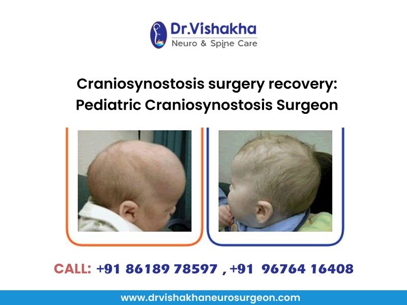
Craniosynostosis surgery recovery: Pediatric Craniosynostosis Surgeon Dr Vishakha Patil explains
Recovery after craniosynostosis surgery is an important process that varies from child to child, but there are some common aspects that parents and caregivers can expect:
Recovery Immediate Post-Surgery
- Hospital Stay: Most children remain in the hospital for a few days following the surgery for monitoring. Healthcare professionals will keep a close eye on vital signs, hydration, and pain management.
- Pain Management: Pain relief is a significant part of recovery. Doctors typically prescribe pain medication to manage discomfort in the initial days after surgery. It’s essential to follow the doctor’s instructions regarding medication dosages and schedules.
- Monitoring for Complications: After surgery, the medical team will watch for potential complications, such as bleeding, infection, or neurological issues. Any signs of unusual swelling, fever, or worsening symptoms should be addressed immediately.
Recovery First Few Days at Home
- Activity Restrictions: For the first couple of weeks, it’s crucial to limit physical activity. This includes avoiding rough play and limiting activities that could put pressure on the head, such as swimming or sports.
- Wound Care: The surgical site will need proper care. Parents are usually given instructions on how to clean and care for the incision area to promote healing while preventing infection. Keeping the area dry and clean is important.
- Follow-Up Appointments: Regular check-ups with the surgeon will be scheduled to monitor the healing process. These appointments are essential to ensure that the skull is healing correctly and that the child’s development is on track.
Recovery Weeks Following Surgery
- Swelling and Bruising: Swelling around the eyes and on the head is common and can take a few weeks to subside. Bruising may also appear, but it typically resolves over time.
- Resuming Normal Activities: Gradually, children can return to their normal activities, but full clearance from the surgeon is essential before resuming more vigorous activities or sports. This may take several weeks to a few months depending on individual healing.
- Emotional Support: Recovery can be a stressful time for both the child and the family. Providing emotional support and reassurance can help the child cope with the changes in their appearance and the experience of surgery.
- Long-Term Follow-Up: Even after physical recovery, ongoing assessments may be needed to monitor head shape and brain development as the child grows. This may include additional imaging studies or consultations with specialists.
Overall, while the recovery period can be challenging, with proper care and monitoring, most children do well and achieve positive outcomes, both in terms of head shape and overall health. Communication with healthcare providers is key to navigating the recovery process effectively.
About Dr Vishakha:
Dr Vishakha Basavraj Karpe is a highly skilled senior consultant at Rainbow Children’s Hospital in Banjara Hills and Hydernagar Hyderabad. She is known for her comprehensive care approach and is one of the few dedicated leading paediatric neurosurgeons in the city and India with over ten years of extensive experience in pediatric neurosurgery. Her expertise includes treating hydrocephalus, spinal dysraphism, craniosynostosis, paediatric brain infections, brain and spine tumours and stroke surgery. She has a special interest in craniosynostosis surgery, which is done only in very few centres in India.
Proficiency of Dr Vishakha:
-
- Hydrocephalus (increased fluid in the brain): The procedure involves an endoscopic third ventriculostomy and CSF diversion (VP shunt) to treat complex hydrocephalus.
- Craniosynostosis (abnormal head shape due to untimely cranial sutures fusion) surgeries: Helmet therapy is a technique that is used in both endoscopic and open surgery.
- Spinal dysraphisms(Spina Bifida)- (spinal abnormalities present by birth) – surgical repair
- Encepahaocles repair surgery.
-
- Vascular conditions and stroke surgeries: revascularization surgeries for moya moya disease.
- Pediatric brain and spine tumour surgeries.
-
- Pediatric brain and spine infection surgeries: Endoscopic and open surgeries for brain and spine infections.
- Pediatric traumatic brain and spinal injury.
- Antenatal counselling for congenital fatal neurosurgical conditions.