Few moments feel as unsettling as reading an unfamiliar term on your child’s scan report. Often, the scan was done for something unrelated, such as a minor fall or a headache, and the word “cyst” suddenly appears in the findings. Questions follow quickly. Is it serious? Will it grow? Does my child need surgery?
The reassuring news is that an arachnoid cyst in children is usually harmless. Many children live with one their whole lives without a single problem. Still, a small number do need treatment, and knowing the difference helps parents stay calm and act at the right time.
Quick Answer Summary
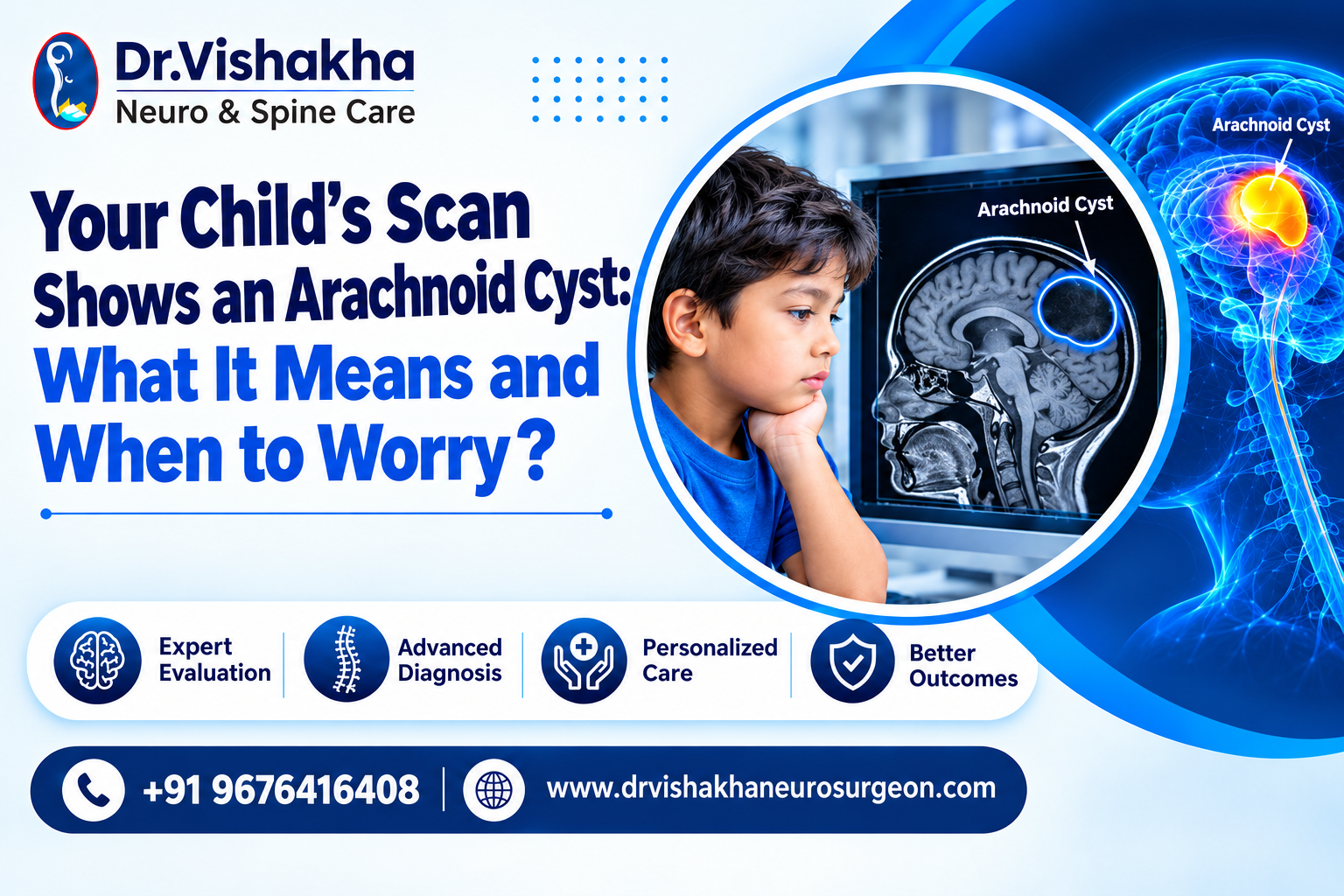
An arachnoid cyst in children is a fluid filled sac in child brain tissue coverings, formed between the brain and the arachnoid membrane. It is not a tumour and is not cancerous. Most cysts are present from birth, cause no symptoms, and are found by chance during a scan. They usually need only regular monitoring. Treatment is considered when a cyst grows, presses on the brain, or causes symptoms such as headaches, seizures, an enlarging head, vomiting, or developmental delay. When needed, surgery is generally safe and highly effective. A pediatric neurosurgeon can assess whether your child’s cyst needs observation or treatment.
What Are Arachnoid Cysts?
The brain is wrapped in three protective layers called the meninges. The middle layer is the arachnoid membrane, named after its delicate, spider-web-like appearance. Between these layers flows cerebrospinal fluid (CSF), the clear liquid that cushions the brain.
Sometimes, during early development in the womb, this membrane splits and forms a pocket. That pocket fills with CSF, creating a cyst. Think of it as a small water pouch sitting beside the brain rather than inside it.
An arachnoid cyst in children is found in roughly 1 to 3 out of every 100 scans. Doctors often call such chance discoveries incidental findings, because they turn up while looking for something else.
There are two broad types:
| Type | How It Forms | How Common |
| Primary (congenital) | Develops before birth due to a split in the arachnoid layer | Most common |
| Secondary (acquired) | Forms later after head injury, infection, bleeding, or brain surgery | Less common |
Arachnoid Cyst Location
Where the cyst sits matters a great deal. Location often decides which symptoms may appear and how treatment is planned.
| Location | Area of the Brain | Possible Effects |
| Middle cranial fossa | Near the temple, beside the Sylvian fissure | Most common site, about half of all cases; often silent |
| Posterior fossa | Back of the head, near the cerebellum | Balance problems, fluid blockage |
| Suprasellar region | Centre of the brain, near the pituitary gland | Vision problems, hormone changes, head bobbing |
| Convexity | Over the surface of the brain | Headaches, seizures |
| Quadrigeminal cistern | Deep, behind the midbrain | Hydrocephalus (fluid build-up) |
Cysts are seen more often in boys and frequently appear on the left side of the brain.
Causes: Why Do These Happen?
In most cases, nothing a parent did or did not do caused the cyst. Primary cysts are a developmental variation, much like a birthmark, that occurs while the baby is forming in the womb.
The exact reason the arachnoid membrane splits is still not fully understood. Some known associations include:
- Developmental changes in the meninges during early pregnancy
- Head injuries, which can lead to secondary cysts
- Brain infections such as meningitis
- Bleeding in the brain or previous brain surgery
- Rare genetic conditions, including certain metabolic disorders
For most families, it simply helps to know that an arachnoid cyst in children is not the result of anything that happened during routine care or play.
Arachnoid Cyst Symptoms
Most children with a cyst have no symptoms at all. Problems arise only when a cyst is large enough to press on nearby brain tissue or block the normal flow of CSF.
Common Brain Cyst in Child Symptoms
Parents should watch for these brain cyst in child symptoms:
- Persistent or worsening headaches, especially in the morning
- Nausea or repeated vomiting
- Seizures
- Unusual sleepiness or irritability
- Problems with balance or coordination
- Blurred or double vision
- Delayed speech or motor milestones
Arachnoid Cyst Brain Toddler Signs
In babies and younger children, the skull is still soft and growing, so signs can look different. With an arachnoid cyst brain toddler presentation, parents may notice:
- A head that grows faster than expected
- A bulging soft spot (fontanelle)
- Asymmetry or a bulge on one side of the skull
- An involuntary bobbing movement of the head (seen with suprasellar cysts)
When to Worry About Arachnoid Cyst Findings
Knowing when to worry about arachnoid cyst results can ease a lot of anxiety. Seek prompt medical review if your child has:
- A severe headache with vomiting
- A new seizure
- Sudden drowsiness or confusion
- A head injury, even a mild one, followed by new symptoms
Note: A cyst that causes no symptoms is usually not an emergency. Sudden changes in behaviour, alertness, or vision always deserve urgent attention.
Risk Factors and Complications of Arachnoid Cysts
Risk Factors
- Being male
- A history of head trauma, brain infection, or bleeding
- Certain inherited or metabolic conditions
Possible Complications
While uncommon, complications can occur:
- Hydrocephalus: Fluid builds up when the cyst blocks CSF pathways.
- Cyst rupture or bleeding: Usually after a blow to the head. This can lead to a fluid or blood collection called a subdural hygroma or haematoma.
- Pressure on brain structures: Can affect vision, hormones, or development depending on location.
Some specialists advise extra care in contact sports for children with large middle fossa cysts. Your neurosurgeon can guide you on what is suitable for your child.
Conditions We Often See (Diagnosis and Tests)
In clinics, arachnoid cysts are often seen alongside or confused with other conditions, such as hydrocephalus, enlarged head size, and other benign fluid spaces. Accurate testing helps tell them apart.
| Test | Purpose |
| MRI scan | The most reliable way to confirm the cyst and see its exact size and position |
| CT scan | Quick assessment, especially after an injury |
| Cine MRI / CSF flow study | Shows whether fluid is flowing normally |
| Eye examination | Checks for pressure on the optic nerves |
| Developmental assessment | Tracks speech, motor skills, and learning |
| Hormone tests | Used when the cyst is near the pituitary gland |
What Happens Next?
Most small, symptom-free cysts only need monitoring. This usually means periodic scans and regular check-ups to make sure the cyst is stable.
When Surgery Is Recommended
Arachnoid cyst surgery in kids is considered when the cyst grows, causes symptoms, or leads to hydrocephalus. Common approaches include:
- Endoscopic fenestration: A minimally invasive procedure where a tiny camera is used to make an opening in the cyst wall, letting fluid drain into normal CSF spaces.
- Microsurgical fenestration: An open procedure through a small opening in the skull for cysts that cannot be reached endoscopically.
- Cyst-peritoneal shunt: A thin tube that drains cyst fluid into the abdomen, used in selected cases.
The outlook after arachnoid cyst surgery in kids is generally excellent. Many children see their symptoms improve within weeks.
Choosing the Right Care
Access to experienced specialists makes a real difference. Specialised pediatric brain cyst treatment India offers today includes advanced endoscopic techniques at dedicated children’s hospitals, bringing world-class care closer to home for families.
Questions to Ask Your Specialist
- Where exactly is the cyst, and how large is it?
- Is it pressing on any part of the brain?
- Does my child need treatment now, or can we monitor it?
- How often will follow-up scans be needed?
- Which symptoms should prompt an urgent visit?
- Are there any sports or activities my child should avoid?
- If surgery is needed, which approach do you recommend, and why?
Frequently Asked Questions
1. Is an arachnoid cyst in children dangerous?
In most cases, no. An arachnoid cyst in children is benign and often causes no symptoms. Only a small percentage need treatment.
2. What are the early brain cyst in child symptoms?
The early brain cyst in child symptoms include frequent headaches, vomiting, seizures, balance problems, and developmental delays. Many children, however, show no symptoms at all.
3. Arachnoid cyst brain toddler findings: what should parents watch for?
When there is an arachnoid cyst brain toddler diagnosis, watch for rapid head growth, a bulging soft spot, irritability, and delayed milestones.
4. When to worry about arachnoid cyst growth?
Parents wondering when to worry about arachnoid cyst changes should look out for new seizures, severe headaches, vomiting, or sudden drowsiness, especially after a head injury.
5. Can a fluid filled sac in a child’s brain go away on its own?
A fluid filled sac in child brain coverings rarely disappears completely, though some cysts shrink over time. Most remain stable and harmless.
6. Is arachnoid cyst surgery in kids safe?
Yes. Arachnoid cyst surgery in kids, especially endoscopic surgery, is considered safe and effective when performed by an experienced pediatric neurosurgeon.
7. Where can I find pediatric brain cyst treatment India families trust?
Leading children’s hospitals with dedicated pediatric neurosurgery teams offer trusted pediatric brain cyst treatment India wide, including in Hyderabad.
Conclusion
Finding a cyst on your child’s scan can feel alarming, but knowledge brings clarity. An arachnoid cyst in children is usually a quiet, harmless finding that needs nothing more than occasional monitoring. When a cyst does cause problems, modern treatment options are safe, effective, and often minimally invasive.
The key is a careful evaluation by a pediatric neurosurgeon who can explain what the scan means for your child specifically, and guide your family with confidence.
Proficiency of Dr Vishakha – Neurosurgeries Expertise
Hydrocephalus (increased fluid in the brain): The procedure involves an endoscopic third ventriculostomy and CSF diversion (VP shunt) to treat complex hydrocephalus.
Craniosynostosis (abnormal head shape due to premature cranial suture fusion) surgeries: Helmet therapy is a technique that is used in both endoscopic and open surgery.
Spinal dysraphisms(Spina Bifida)– (spinal abnormalities present by birth) – surgical repair
Encephalocele repair surgery.
Vascular conditions and stroke surgeries: revascularisation surgeries for moyamoya disease.
Pediatric brain and spine tumour surgeries.
Pediatric brain and spine infection surgeries: Endoscopic and open surgeries for brain and spine infections.
Pediatric traumatic brain and spinal injury.
Antenatal counselling for congenital fatal neurosurgical conditions.
Related Links
Related treatment pages:
- Craniosynostosis: https://drvishakhaneurosurgeon.com/craniosynostosis/
- Pediatric Neurosurgery: https://drvishakhaneurosurgeon.com/pediatric-neurosurgery/
- Hydrocephalus: https://drvishakhaneurosurgeon.com/hydrocephalus/
- Antenatal Counselling: https://drvishakhaneurosurgeon.com/antenatal-counselling/
Other blog posts:
- Spina Bifida Journey: From Antenatal Diagnosis to Postnatal Repair: https://drvishakhaneurosurgeon.com/spina-bifida-journey-from-antenatal-diagnosis-to-postnatal-repair/
- Understanding Your Baby’s Milestones: How Brain & Spine Health Plays a Crucial Role: https://drvishakhaneurosurgeon.com/understanding-your-babys-milestones-how-brain-spine-health-plays-a-crucial-role-dr-vishakha/
- Is Your Baby’s Soft Spot Normal? Here’s How to Tell: https://drvishakhaneurosurgeon.com/is-your-babys-soft-spot-normal-heres-how-to-tell-dr-vishakha-explains/
Book a Consultation
If your child’s scan has shown an arachnoid cyst, or if you have noticed any of the warning signs above, an expert opinion can bring clarity and peace of mind.
Book a consultation with Dr. Vishakha Karpe today — call +91 8618978597 or +91 9676416408, or visit drvishakhaneurosurgeon.com to schedule your visit.