
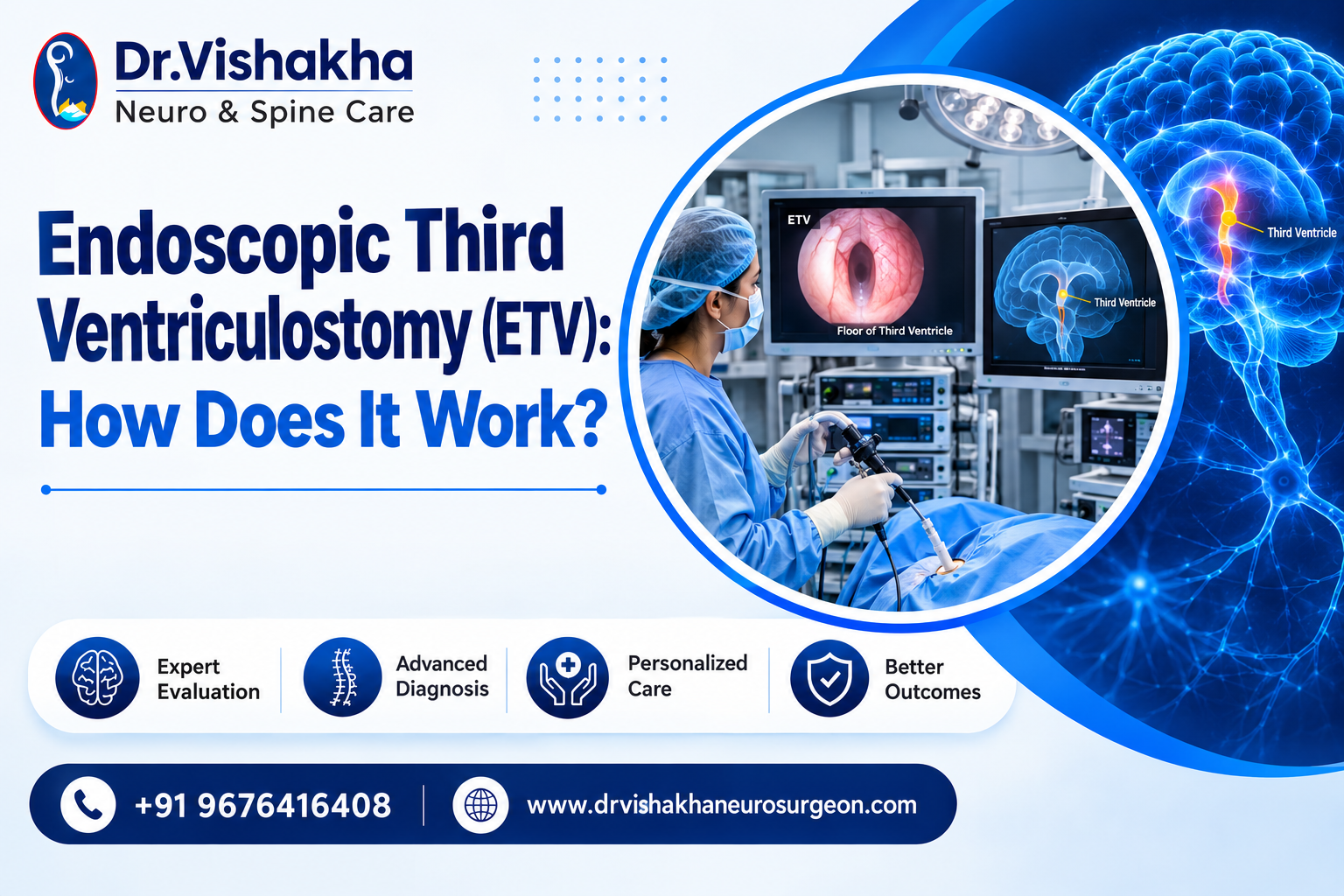
Endoscopic Third Ventriculostomy (ETV) is a minimally invasive neurosurgical procedure used to treat hydrocephalus, a condition caused by the abnormal accumulation of cerebrospinal fluid (CSF) within the brain. Instead of implanting an artificial drainage tube (shunt), the neurosurgeon uses a camera-guided endoscope to create a small opening in the floor of the third ventricle, allowing CSF to resume its natural flow. The ETV procedure typically takes under an hour and generally involves a shorter recovery period than shunt surgery. For eligible patients with obstructive hydrocephalus, it offers a safe and effective alternative to lifelong shunt dependence.
Understanding What Endoscopic Third Ventriculostomy Really Is
To understand how ETV works, it helps first to understand what goes wrong in hydrocephalus. Cerebrospinal fluid circulates continuously through a series of connected chambers in the brain, cushioning and nourishing neural tissue before draining and being reabsorbed. When this pathway is obstructed, whether by a congenital narrowing, a growth, or scarring from infection, fluid accumulates, and pressure inside the skull begins to rise.
Endoscopic Third Ventriculostomy addresses this at its source. Using a slender endoscope fitted with a camera and light source, the surgeon navigates through the brain’s existing ventricular pathways and creates a precise opening in the floor of the third ventricle. This opening establishes a new, direct route for CSF to reach the subarachnoid space, where it is naturally reabsorbed into the bloodstream.
The result is a physiological solution rather than a mechanical one. No implanted hardware, no external tubing, just a restored pathway that allows the brain’s own fluid dynamics to function as intended.
How ETV Helps With Hydrocephalus
Hydrocephalus is not a single, uniform diagnosis. It varies depending on where the CSF pathway is obstructed, and this location determines whether ETV is a suitable treatment.
When the blockage occurs within reach of the endoscope, typically at or near the third or fourth ventricle, ETV for hydrocephalus becomes a highly effective option. By creating a bypass at the site of obstruction, the procedure restores CSF circulation through a natural route, rather than diverting it through an external device that requires ongoing monitoring.
This distinction matters clinically. ETV treatment is specifically suited to obstructive hydrocephalus, where the issue is a physical blockage in flow. It is generally not the first choice for communicating hydrocephalus, where the problem lies in fluid absorption rather than flow obstruction. This is why detailed imaging, usually an MRI and careful clinical evaluation are essential before recommending the procedure.
Why Do These Happen?
Understanding the underlying cause of hydrocephalus helps clarify why treatment decisions vary from patient to patient. Common causes include:
- Congenital anomalies — some infants are born with a narrowed or malformed CSF pathway (such as aqueductal stenosis), restricting flow from birth
- Tumours or cysts — growths within or near the ventricular system can physically obstruct circulation
- Infections — conditions such as meningitis can cause scarring that blocks normal CSF drainage
- Intraventricular haemorrhage — bleeding, particularly in premature infants, can obstruct the delicate ventricular channels
- Traumatic injury — head trauma can disrupt normal fluid dynamics within the brain
Regardless of the underlying cause, the outcome is the same: CSF has nowhere to go, pressure builds within the skull, and if left untreated, this can affect a child’s neurological development, vision, motor coordination, and cognitive function.
Importance of ETV in Functional Neurosurgery
Within the field of functional neurosurgery, ETV occupies a distinct and important role. Rather than replacing a disrupted physiological process with a permanent mechanical device, it restores the brain’s own fluid pathways.
This distinction carries particular weight for pediatric patients. A shunt requires long-term monitoring, may need surgical revision as a child grows, and carries a lifelong risk of infection or malfunction. ETV surgery, when clinically appropriate, avoids many of these long-term considerations by working within the body’s existing anatomy rather than around it.
For this reason, neurosurgeons regard ETV as a cornerstone technique in modern hydrocephalus management, one that reflects broader advances in endoscopic neurosurgery, where large incisions have given way to precise, millimetre-scale interventions.
Conditions We Often See
In clinical practice, certain presentations of hydrocephalus recur consistently:
| Condition | How ETV Helps |
| Aqueductal stenosis | Bypasses the narrowed channel entirely |
| Post-infective hydrocephalus | Creates a new route around scarred tissue |
| Tumour-related obstruction | Relieves pressure while further tumour management is planned |
| Chiari malformation with hydrocephalus | Reduces fluid buildup alongside corrective surgery |
| Failed shunt cases | Offers a shunt-free alternative for select patients |
Each case, however, depends on individual anatomy and the precise nature of the obstruction. This is why imaging review and specialist consultation remain essential before finalising a treatment plan.
What Happens Next?
Recovery following ETV surgery is generally more straightforward than many patients anticipate. Most patients remain in the hospital for a few days for observation, and many return to normal activity within one to two weeks. Follow-up MRI scans are used to confirm that the newly created pathway remains open and functioning as intended.
It is important to note that ETV surgery side effects can occur, including headache, low-grade fever, or temporary CSF leakage. In a subset of cases, the created opening may narrow over time, which can require a repeat procedure. For this reason, long-term follow-up is not optional, it is a core part of post-operative care.
Compared to a permanent ETV shunt alternative, ETV generally results in fewer lifetime interventions for appropriately selected patients, though outcomes vary based on individual anatomy and the underlying cause of hydrocephalus.
Questions to Ask Your Specialist
A neurosurgical consultation can raise many questions. The following are worth discussing directly with your specialist:
- Is this hydrocephalus obstructive in nature, and therefore suitable for ETV?
- What do the imaging findings show, and how do they support this recommendation?
- What is the realistic success rate for a case with this specific anatomy?
- What symptoms after surgery would require immediate medical attention?
- How many ETV procedures has the surgeon performed?
A qualified specialist will welcome these questions as part of informed decision-making.
FAQs
- Is Endoscopic Third Ventriculostomy painful?
The procedure is performed under general anesthesia, so no pain is experienced during surgery. Mild post-operative headache is common and is typically managed with medication. - How long does the ETV procedure take?
Most procedures are completed within 45 minutes to an hour, depending on individual anatomy. - Is ETV a permanent solution for hydrocephalus?
For many patients, the created pathway remains functional long-term. In some cases, a repeat procedure or a shunt may later be required if the opening narrows. - What is the difference between ETV and a shunt?
A shunt uses a permanently implanted tube to drain CSF externally. ETV creates an internal pathway using the body’s own anatomy, without implanted hardware. - Can adults undergo ETV, or is it limited to children?
Both children and adults may be candidates, depending on the type and underlying cause of hydrocephalus. - What are the risks associated with ETV surgery?
Risks are generally low but may include infection, bleeding, transient memory disturbance, or closure of the created opening requiring further intervention.
Conclusion
A diagnosis of hydrocephalus can be overwhelming, particularly for families navigating treatment decisions for a child. Endoscopic Third Ventriculostomy has meaningfully changed the treatment landscape, offering smaller incisions, shorter recovery periods, and for eligible patients, freedom from lifelong shunt dependence. Understanding the fundamentals of etv hydrocephalus treatment allows families to engage confidently with their specialist and make informed decisions about care.
Proficiency of Dr Vishakha – Neurosurgeries Expertise
Hydrocephalus (increased fluid in the brain): The procedure involves an endoscopic third ventriculostomy and CSF diversion (VP shunt) to treat complex hydrocephalus.
Craniosynostosis (abnormal head shape due to premature cranial suture fusion) surgeries: Helmet therapy is a technique that is used in both endoscopic and open surgery.
Spinal dysraphisms(Spina Bifida)– (spinal abnormalities present by birth) – surgical repair
Encephalocele repair surgery.
Vascular conditions and stroke surgeries: revascularisation surgeries for moyamoya disease.
Pediatric brain and spine tumour surgeries.
Pediatric brain and spine infection surgeries: Endoscopic and open surgeries for brain and spine infections.
Pediatric traumatic brain and spinal injury.
Antenatal counselling for congenital fatal neurosurgical conditions.
Related Links
Related treatment pages:
- Craniosynostosis: https://drvishakhaneurosurgeon.com/craniosynostosis/
- Pediatric Neurosurgery: https://drvishakhaneurosurgeon.com/pediatric-neurosurgery/
- Hydrocephalus: https://drvishakhaneurosurgeon.com/hydrocephalus/
- Antenatal Counselling: https://drvishakhaneurosurgeon.com/antenatal-counselling/
Other blog posts:
- Spina Bifida Journey: From Antenatal Diagnosis to Postnatal Repair: https://drvishakhaneurosurgeon.com/spina-bifida-journey-from-antenatal-diagnosis-to-postnatal-repair/
- Understanding Your Baby’s Milestones: How Brain & Spine Health Plays a Crucial Role: https://drvishakhaneurosurgeon.com/understanding-your-babys-milestones-how-brain-spine-health-plays-a-crucial-role-dr-vishakha/
- Is Your Baby’s Soft Spot Normal? Here’s How to Tell: https://drvishakhaneurosurgeon.com/is-your-babys-soft-spot-normal-heres-how-to-tell-dr-vishakha-explains/
Book a Consultation
If your child has been diagnosed with hydrocephalus, or you would like a specialist second opinion, timely consultation is important.
Book a consultation with Dr. Vishakha Karpe today — call +91 8618978597 or +91 9676416408, or visit drvishakhaneurosurgeon.com to schedule your visit.